Fig. 3.
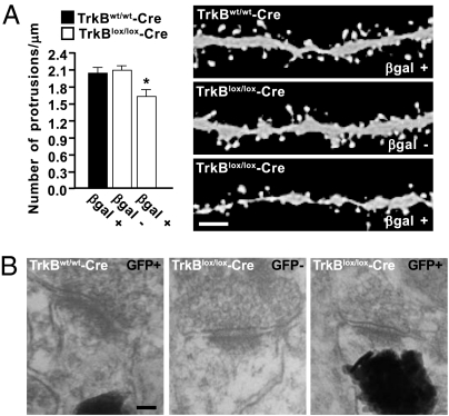
Spine density is regulated by TrkB in adult-born neurons. (A) Graphs depict the quantification of protrusions in dendritic segments of newborn neurons transduced with retrovirus expressing GFP in tamoxifen-induced TrkBlox/lox-Cre R26R (n = 24 dendritic segments; 18 βgal+; 11 βgal−; 3 animals) and control mice (n = 60 dendritic segments; 26 βgal+; 3 animals) mice. The density of protrusions is expressed as the number of protrusions per micrometer of dendritic length (*, P < 0.01). Representative images show spine morphology in reporter-positive (βgal+) or -negative (βgal−) cells 28 dpi. (Scale bar, 2 μm.) (B) Representative electron micrographs showing synaptic contacts of newborn neurons in tamoxifen-induced TrkBlox/lox-Cre Z/EG and control mice. Reporter-positive cells (GFP+) show GFP fluorescence as photoconverted electron-dense signals at postsynaptic sites. A synapse from a reporter-negative cell (GFP−) is shown as a reference. (Scale bar, 50 nm.)

