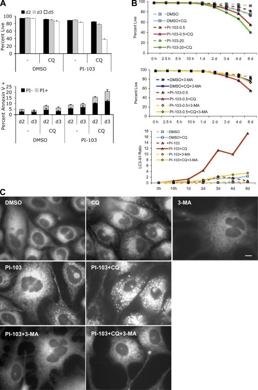
Figure 5.
CQ accelerated cell death in combination with PI-103. (A) PC3 cells were treated with DMSO or 0.5 μM PI-103 in the presence or absence of 10 μM CQ under 0.5% FBS. Cell viability was determined by PI exclusion at days 2, 3, and 5. Annexin V staining was analyzed at days 2 and 3 and broken down into PI+ or PI− populations. (B) Time course of cell viability in PC3 cells treated with 0.5 (PI-103-0.5) or 20 μM (PI-103-20) PI-103 with or without 10 μM CQ or 3 mM 3-MA. PC3 cells pretreated with PI-103 for 24 h under 1% FBS were split into medium containing 0.5% FBS in the presence or absence of CQ. 3-MA was added immediately before PI-103, 24 h before CQ addition. Cell viability was determined by PI exclusion at the indicated time points after CQ addition. Error bars represent SEM (n = 3). LC3-II to LC3-I ratios were determined from quantitation of immunoblots (with 0.5 μM PI = 103. (C) CQ dramatically increased the size and number of MDC+ vacuoles in PC3 cells treated with PI-103, whereas 3-MA suppressed this effect. Cells were cultured in medium containing 0.5% FBS and treated with DMSO, 0.5 μM PI-103, 10 μM CQ, and 5 mM 3-MA, alone or in combinations as indicated. MDC staining at 48 h is shown. Bar, 10 μm.

