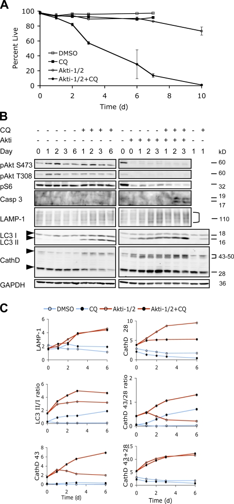
Figure 6.
CQ accelerated cell death in combination with Akti-1/2. (A) PC3 cells were treated with DMSO or 4 μM Akti-1/2 in the presence or absence of 10 μM CQ under 0.5% FBS. Cell viability was determined by PI exclusion over the course of 10 d. Error bars represent SEM. Representative data from one of three independent experiments are shown. (B) Immunoblot analysis of cell lysates collected at the indicated time points from the experiment shown in A. Arrowheads indicate the positions for LC3-I and -II, CathD 43, and CathD 28. Quantifications of the indicated markers are shown in C. CathD 43, the 43–50-kD forms of cathepsin D precursors. CathD 28, the 28-kD cathepsin D heavy chain.

