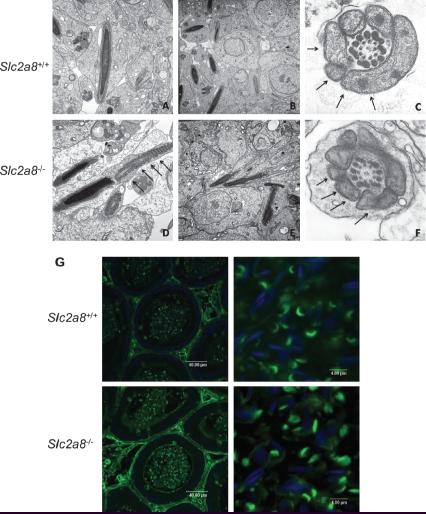
Figure 6.
Electron microscopy of testis of Slc2a8+/+ (A–C) and Slc2a8−/− mice (D–F) and analysis of cauda epididymal sperm with Pisum sativum agglutinine. (A) Early spermatides, (B, D, E) late spermatides, (C, F) cross section of a late spermatide, cut in the mid-piece region of the tail. Arrows depict the mitochondria (magnification approx. A and D × 18,000; B and E × 12,000; C and F × 40,000). (G) Sections of the cauda epididymis of Slc2a8+/+ and Slc2a8−/− males at the age of 10–12 weeks were stained with Pisum sativum agglutinine and analyzed by confocal laser scanning microscopy. Nuclei were co-stained with DAPI.

