
Figure 5.
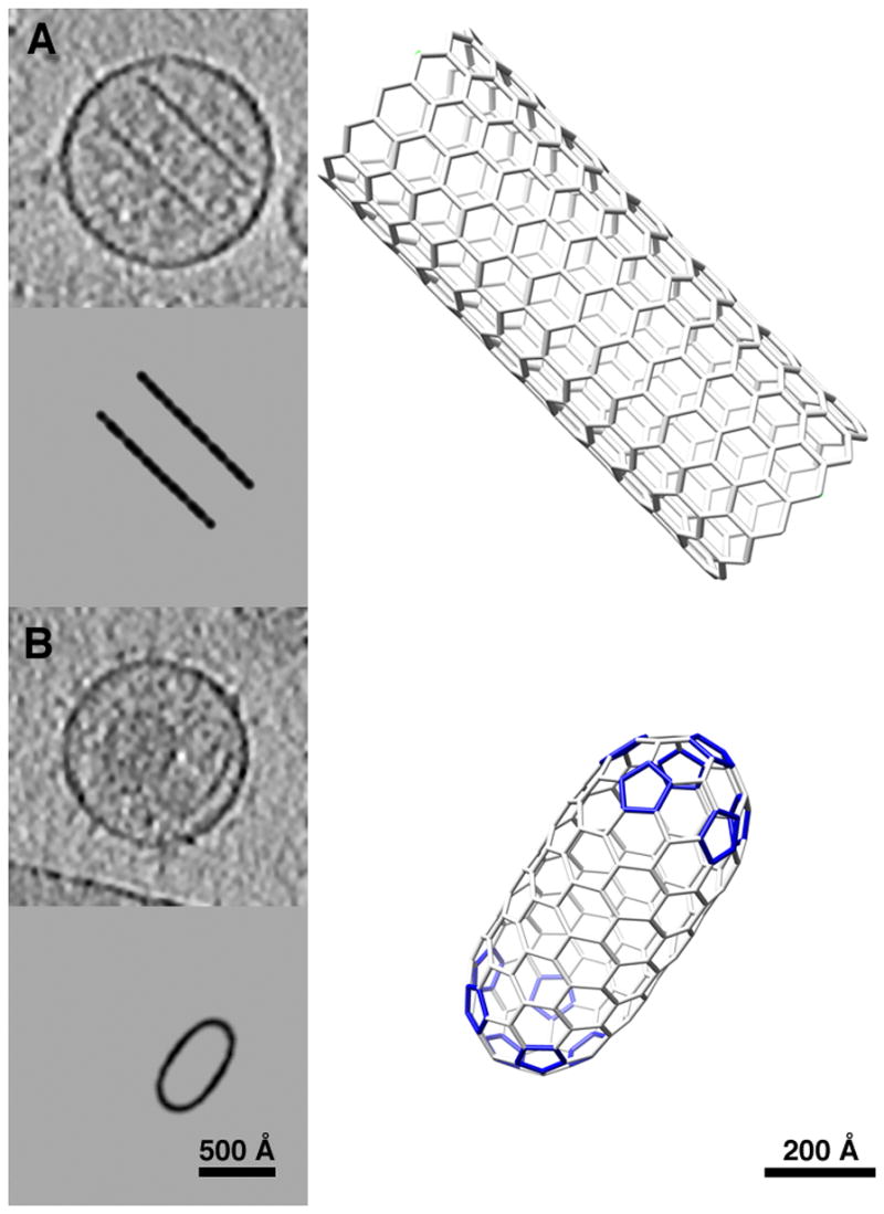
Polyhedral models similar to (A) open and (B) closed (“lozenge”) tubular cores observed in tomograms of RSV virions. In each panel a slice through the tomogram is shown in the upper left, with a corresponding slice through the modeled core beneath, and a 3D representation to the right.