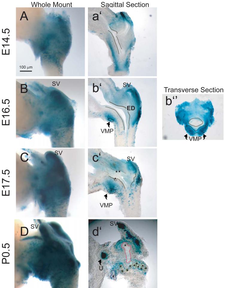
Figure 1. Temporal and spatial expression of retinoid activity in the UGS of male Tg(RARE-Hspa1b/lacZ)12Jrt transgenic mouse fetuses.
To assess the temporal expression of retinoid signaling during prostatic budding initiation and elongation, the UGS from fetal male mice that express a LacZ transgene under control of a RARE-containing promoter were collected at regular intervals during development from embryonic day (E)14.5 to postnatal day (P)0.5 and probed for β-galactosidase activity. Photographs are representative of n = 5 UGS specimens per developmental time point. To evaluate global retinoid activity and activity within specific UGS structures, β-galactosidase activity in UGS specimens was visualized by whole mount imaging (A-D), imaging of sections cut near the mid-sagittal plane (a’-c’) or along the sagittal plane near the lateral surface (d’), and imaging of sections taken along the transverse plane (b”). To enhance visualization, a dotted black line was added to illustrate the boundary between the inner UGS epithelial cell layer and outer mesenchymal cell layer and a solid black line was added to indicate the ventral mesenchymal pad (VMP). Prostatic buds in tissue sections are indicated by asterisks and by colored dotted lines in whole-mount images (blue = ventral buds, green = dorsolateral buds, and red = anterior buds). SV: seminal vesicle; ED: ejactulatory duct; U: ureter.

