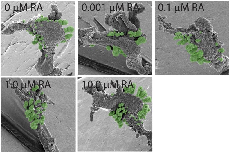
Figure 6. Prostatic bud formation in male mouse UGS organ cultures exposed to RA.
UGS from E14.5 male C57BL/6J mouse fetuses were incubated for 3 d in organ culture media containing vehicle or graded concentrations of RA (0-10 μM). Media and RA were replenished after each day. At the end of the incubation, UGE was separated from UGM as described in methods and visualized by SEM. A micrograph representing a single UGE from each exposure group is shown (results are n ≥ 6 independent samples per group from at least 3 separate litters). Prostatic buds are pseudocolored green.

