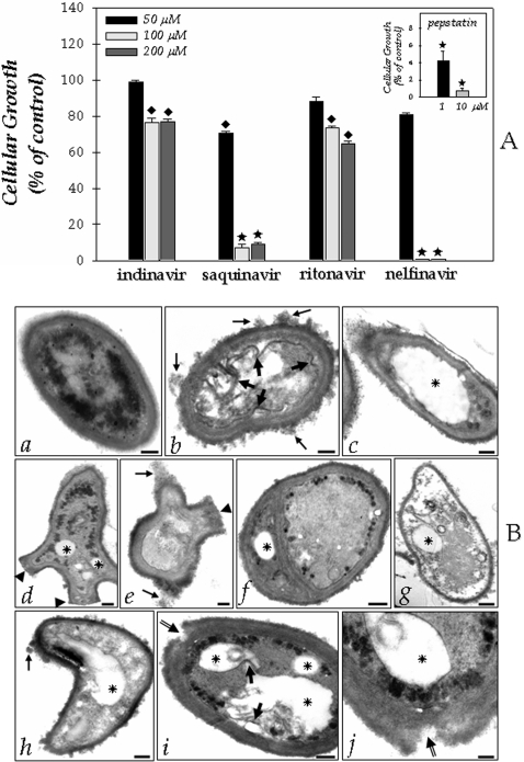
Figure 2. Functional implications of HIV PIs on the Fonsecaea pedrosoi conidial cells development.
(A) Effect of aspartyl PIs on the F. pedrosoi conidial growth. After growth in Czapek medium for 5 days at room temperature, the conidia were harvested, washed with PBS and 1×10³ cells were incubated for 1 hour in the absence or in the presence of different concentrations of the following PIs: indinavir, saquinavir, ritonavir, nelfinavir (50, 100 and 200 µM) and pepstatin A (1 and 10 µM). After that, the conidial cells were washed in PBS and inoculated in a fresh solid Czapek medium to measure the CFU. The control (435.2±12.2 CFU) was taken as 100%. The values represent the mean±standard deviation of three independent experiments performed in triplicate. Symbols denote systems treated with PIs that had a growth rate significantly different from the control (♦, P<0.05; ★, P<0.001; Student's t test). (B) Effect of aspartyl PIs on the F. pedrosoi ultrastructure. After growth in Czapek medium for 5 days at room temperature, the conidia were harvested, washed with PBS and 1×10³ cells were incubated for 1 hour in the absence (a) or in the presence of nelfinavir (b–g) or saquinavir (h–j) at 100 µM. Subsequently, the cells were processed by transmission electron microscopy as described in the Experimental Procedures section. Control conidial cells present a characteristic dense cytoplasm with a distinct and compact cell wall (a). Large arrows in (b) and (i) show the invaginations of cytoplasmic membrane. Tiny arrows in (b), (e) and (h) indicate the disorder and detachment of cell wall. Arrowheads in (d) and (e) demonstrate the abnormal cellular division. Asterisks show the enlarged cytoplasmic vacuoles. Double arrows in (i) and (j) appoint the breakage of cell wall starting from extracellular medium. Note in (g) a typical necrotic conidial cell. Scale bars: (a–i), 0.6 µm; (j), 0.15 µm.

