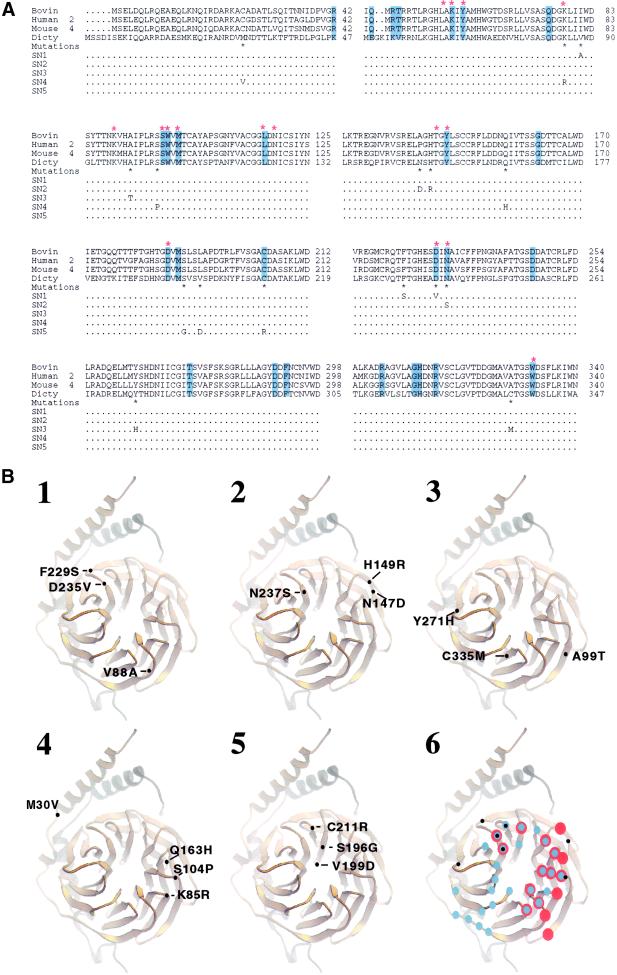
Figure 7.
(A) Amino acid sequence alignment of four Gβ subunits and the substitutions in the SN Gβ subunits. Residues that contact phosducin are highlighted in blue. Red-stared residues interact with Gα. Black-starred residues are substituted in the SN Gβ subunits, and the substituted amino acids in SN1–SN5 are indicated. (B) Ribbon diagram showing the positions of the substitutions in each of the SN Gβ subunits. Gβ is gold; Gγ is silver. The numbers are placed directly on the ribbons according to the Gβ of D. discoideum (A), and positions are placed to the corresponding positions of the transducin Gβγ subunit (Sondek et al., 1996). (1) SN1; (2) SN2; (3) SN3; (4) SN4; (5) SN5. (6) Corresponding residues that contact Gα (red dots) and phosducin (blue dots) and the positions of the substitutions (black dots) that are predicated to cause defects in SN Gβ subunits.

