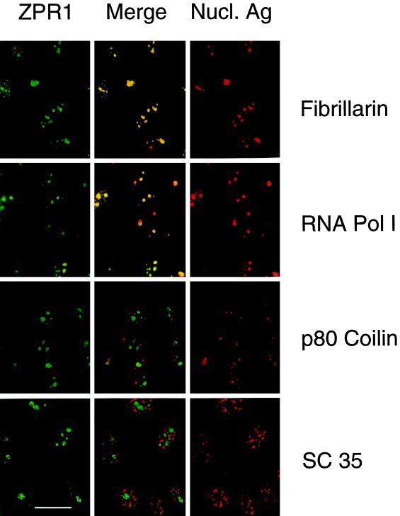
Figure 2.
Nuclear ZPR1 accumulates in the nucleolus. Activated HEp-2 cells were examined by laser scanning confocal microscopy using antibodies to ZPR1 (FITC, green) and nuclear antigens (rhodamine, red). In the merged image, fibrillarin and RNA Pol I show extensive colocalization (yellow), whereas p80 coilin and SC35 splicing factor show extensive segregation (green and red). Bar, 35 μm. Fibrillarin and RNA Pol I are markers for the nucleolus.

