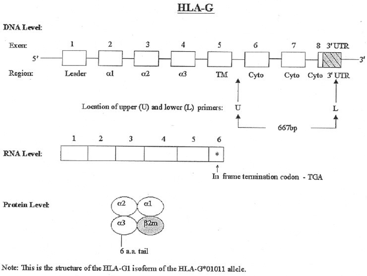
Figure 1.
Structure of HLA-G at the DNA, RNA, and protein levels. At the DNA level, the exons are open boxes and the 3 ′UTR is a shaded, hatched box. The primary RNA transcript is larger than shown; the RNA transcript is shown only up to the stop codon in exon 6. Also shown is the location of the upper (U) and lower (L) primers used in this study.

