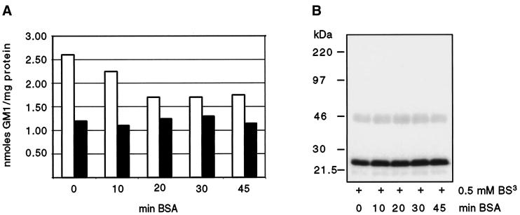
Figure 3.
Incorporation of GM1 into the plasma membrane of MDCK GH-DAF cells. (A) MDCK GH-DAF cells were incubated with 1 μCi/ml tritiated GM1 (final ganglioside concentration 100 μM) for 1 h at 37°C and subsequently washed with BSA for 0, 10, 20, 30, or 45 min (open bars). The additive effect of trypsin treatment (0.1% trypsin for 5 min at 37°C) after washing with BSA is shown as solid bars for each time point. (B) Cross-linking was performed after incubating GM1-loaded cells with BSA for 0, 10, 20, 30, or 45 min. SDS-PAGE, Western blotting, and detection were as described in Figure 1.

