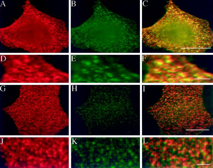
Figure 8.
Inhibition of copatching of influenza HA and GH-DAF by gangliosides. MDCK GH-DAF cells were infected with influenza HA virus and then incubated with DMEM (A–F) or 100 μM GM1 in DMEM (G–L) for 1 h at 37°C. After subsequent treatment with a mixture of monoclonal anti-HA and sheep polyclonal anti-GH antibodies at 4°C, patching was detected using Cy-3-anti-sheep–labeled (red) and FITC-anti-mouse–labeled (green) secondary antibodies. Panels in the left column show distribution of GH, panels in the middle column show distribution of HA, and panels in the right column show the merge of both signals. D–F display a detail of A–C correspondingly, whereas J–L display that of G–I. Bars: F, L, 2 μm; C, I, 8 μm.

