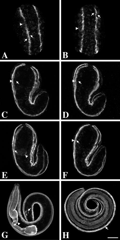
Figure 3.
Localization of UNC-52 isoforms during embryonic development. Embryos were visualized by confocal microscopy. Wild-type embryos were labeled with GM1 (domain III; A, C, E, and G) and GM3 (domain IV; B, D, F, and H). A and B show the dorsal view of a comma-stage embryo (∼350 min); these images are projections of the dorsal Z-sections. Arrows indicate positions of two adjacent muscle cells; the arrowhead indicates intracellular staining. Subsequent images are projections of the complete Z-series. C and D show the lateral view of late comma-stage embryo (350–400 min). Arrows indicate the position of a muscle cell, whereas the arrowheads indicate the basal face of the cell. E and F show the lateral view of a 1.5-fold embryo (∼420 min). The arrow in both panels indicates the basal face of the muscle cells in a dorsal quadrant. Note the pharyngeal staining (arrowhead) in E. G and H show a threefold embryo (∼800 min). Note that GM1 stains the pharynx (arrowhead in G), body-wall muscles, and anal muscles (arrow in G), whereas GM3 only stains the body-wall muscles. Bar, 10 μm.

