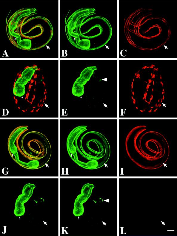
Figure 5.
unc-52(st560) mutant embryos have tissue-specific staining defects and lack a subset of UNC-52 isoforms. Embryos were visualized by confocal microscopy. Embryos in A–F were double-labeled with GM1 (green), which recognizes all UNC-52 isoforms, and DM5.6 (red), which recognizes myosin heavy chain A (MHC A). Small arrows indicate the pharynx, and large arrows indicate a body-wall muscle quadrant. A and D show both channels simultaneously, whereas B, C, E, and F show single-channel images. A–C show a wild-type embryo, whereas D–F show an arrested unc-52(st560) mutant embryo. Note the reduced staining of body-wall muscles with GM1 and disorganization of MHC A in the mutant (E and F; compare with B and C). The pharynx and anal muscles (arrowhead in E) in the mutant, however, exhibit a wild-type staining pattern (compare B and E). Embryos in G–L were double-labeled with GM1 (green, FITC) and MH3 (red, TRSC), which recognizes an epitope in domain IV of UNC-52. G–I show a wild-type embryo; J–L show a unc-52(st560) mutant embryo. G and J show both channels simultaneously. Note the absence of MH3 staining in the mutant embryo in L. Although the unc-52(st560) mutant embryos shown are arrested at the twofold stage, they are comparable to threefold wild-type embryos. Bar, 10 μm.

