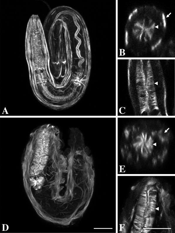
Figure 6.
Phalloidin staining in wild-type and unc-52(null) embryos. Embryos were visualized by confocal microscopy. Wild-type (A–C) and unc-52(st549) (D–F) embryos were labeled with FITC-phalloidin. Images in A and D show staining from all focal planes. Computer-generated cross sections (B and E) and single focal plane images (C and F) are also shown. The arrowhead indicates the basal surface of the pharynx, whereas the arrow indicates the basal face of the body-wall muscles. Note the well organized thin filaments extending from the basal and apical faces of the pharynx in both wild-type and mutant embryos. Also note that actin in the body-wall muscles of the mutant is not associated with the basal cell membrane. Bar, 10 μm.

