Figure 5.
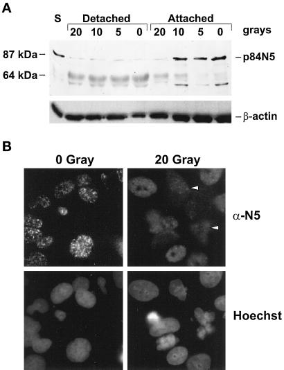
The structure and subcellular localization of endogenous p84N5 is altered during apoptosis. (A) SAOS-2 cells were treated with the indicated dose of ionizing radiation or staurosporine (S). Attached or detached cells were harvested separately 72 h (irradiated) or 12 h (staurosporine) later, and p84N5 expression was analyzed in each sample by Western blotting as in Figure 1D. The positions of p84N5, β-actin, or a 64-kDa molecular weight standard are indicated on the left. (B) SAOS-2 cells were irradiated with the indicated dose of γ-radiation. Three days later, cells were processed for immunofluorescent staining using the α-N5 5E10 monoclonal antibody as described in MATERIALS AND METHODS. Cells were photographed at 630×. The arrows indicate two examples of cytoplasmic N5 staining in cells with pyknotic nuclear morphology. Note the more homogeneous α-N5 staining in the irradiated sample.

