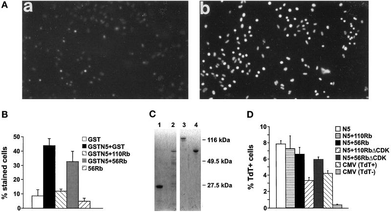
Figure 6.
Association with Rb inhibits p84N5-induced apoptosis. (A) SAOS-2 cells were injected with purified GST (a) or GSTN5 (b). Ninety minutes later, Hoechst dye was added to the media at 1 μg/ml for 10 min before photography under fluorescence microscopy at 100×. All cells within the field of view have been injected. (B) Cells were injected and analyzed as above with the indicated proteins. The percentage of injected cells with apoptotic morphology and bright Hoechst staining was then counted. The results are the mean of at least three experiments. (C) Aliquots (5 μl) of GST (lane 1), GSTN5 (lane 2), p110Rb (lane 3), or p56Rb (lane 4) used for microinjection were resolved by SDS-PAGE and stained with coomassie blue. The position of molecular weight standards is indicated at the right. (D) SAOS-2 cells were transfected with the indicated expression plasmids and analyzed for apoptosis by TUNEL and flow cytometry as in Figure 2C. The percentage of cells containing fragmented DNA is shown as the mean of three experiments. The maximum transfection efficiency under the conditions used was ∼10%, indicating that most of the N5-transfected cells contained fragmented DNA.

