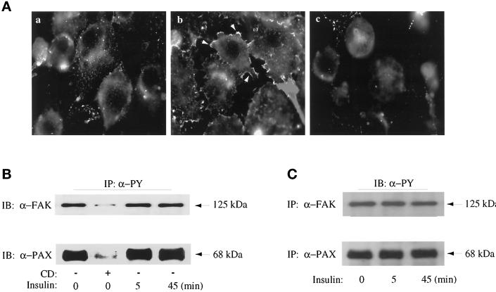
Figure 1.
Effect of insulin and cytochalasin D on the actin cytoskeleton and tyrosine phosphorylation of FAK and paxillin in 3T3-L1 adipocytes. (A) 3T3-L1 adipocytes were grown and differentiated on glass coverslips. The cells were serum deprived for 3 h with no other additions (a), with insulin treatment for the last 5 min (b), or with treatment with 2 μM cytochalasin D during the entire 3 h (c). All cells were fixed and detergent permeabilized, and the actin filaments were stained with rhodamine-conjugated phalloidin, as indicated in MATERIALS AND METHODS. (B) 3T3-L1 adipocytes grown in 10-cm dishes were serum deprived for 3 h in the absence (−) or presence (+) of cytochalasin D. Insulin treatments (100 nM) were for the times indicated and were timed so that their total serum deprivation time ended at 3 h. Cell lysates were prepared and immunoprecipitated with anti-PY conjugated to agarose beads (IP, α-PY), as indicated in MATERIALS AND METHODS. Samples were immunoblotted (IB) anti-FAK (α-FAK) and anti-paxillin (α-PAX) as indicated. (C) 3T3-L1 adipocytes were treated with insulin (100 nM) for the times indicated. Cell lysates were prepared and immunoprecipitated with α-FAK or α-PAX antibodies as described in MATERIALS AND METHODS and then immunoblotted with α-PY as indicated. Shown is one experiment representative of two.

