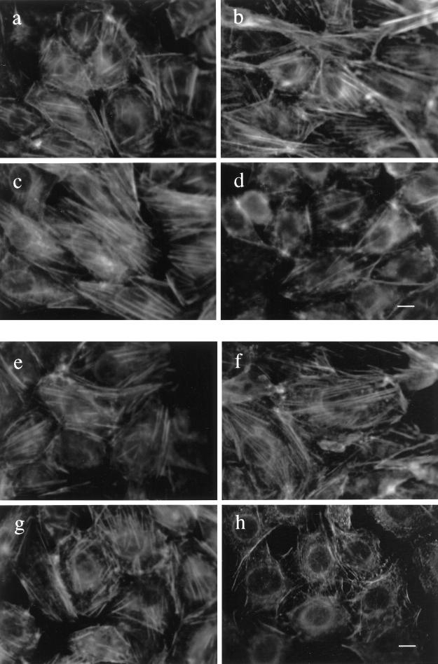
Figure 6.
Subcellular organization of actin fibers in CHO and HTC cells. Confluent cultures of CHO-WT (a and b) and CHO-IR (c and d) cells were serum deprived for 3 h and treated without (a or c) or with 100 nM insulin (b and d) for 15 min. The cells were fixed with 4% paraformaldehyde in PBS for 20 min at room temperature and then permeabilized with 0.2% Triton X-100 in PBS for 20 min. The cells were incubated with rhodamine-conjugated phalloidin (4 U/ml) in PBS for 30 min. The stained actin filaments were examined by fluorescence microscopy. HTC-WT (e and f) and HTC-IR cells (g and h) were treated without (e and g) or with 100 nM insulin (f and h) for 15 min at the end of 3 h of serum starvation and then processed as above. Shown is one experiment representative of three. Bar, 10 μm.

