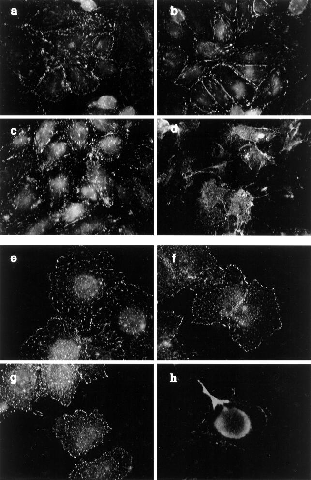
Figure 9.
Subcellular distribution of phosphotyrosine proteins in CHO and HTC cells. Confluent cultures of CHO-WT (a and b) and CHO-IR (c and d) cells were serum deprived for 3 h and treated without (a and c) or with 100 nM insulin (b and d) for 15 min. The cells were fixed, permeabilized, and blocked as described in Figure 6. The cells were then incubated with anti-PY antibody (1:1000) in goat serum-PBS for 60 min. Primary antibody binding was detected with goat anti-mouse antibody conjugated to fluorescein and then examined by fluorescence microscopy. HTC-WT (e and f) and HTC-IR cells (g and h) were treated without (e and g) or with 100 nM insulin (f and h) for 15 min at the end of a 3-h period of serum starvation and then processed as above. Shown is one experiment representative of three.

