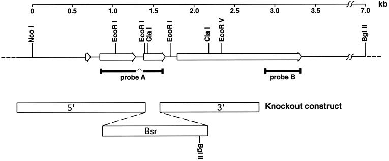
Figure 2.
Genomic organization of the darA gene locus. Probe A was obtained by RT-PCR using oligonucleotides designed from peptide sequences obtained from purified darlin protein. This probe was used to obtain genomic and cDNA clones that encompassed the entire locus. Comparison of the sequence of these clones revealed that the darA open reading frame (open arrows) is interrupted by three introns as indicated. A knockout construct was designed using the indicated portions of the darA gene (5′ and 3′ open bars) and a blasticidin-resistance selectable marker (Bsr). When this construct recombined into the darA locus by a double crossover, it introduced a BglII site 5.5 kb away from the BglII site in the 3′ flanking region of the darA gene (see Figure 7A). Probe B is a 450-bp PCR product from the 3′ end of the open reading frame. It does not overlap with portions included in the knockout construct.

