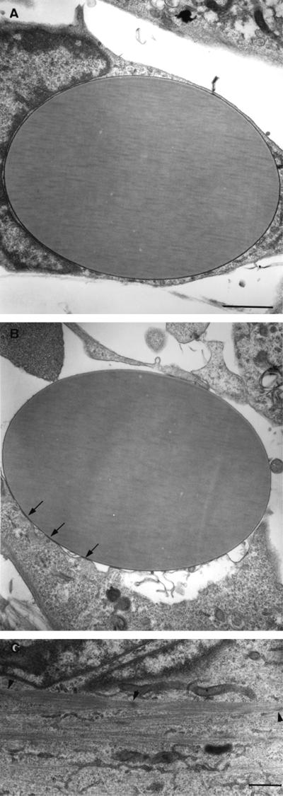
Figure 1.
Transmission electron micrographs of C2 myoblasts treated with beads coated with BSA (A) or NEC (B and C). The cells were incubated with the beads for 48 h in growth medium, fixed, and processed for transmission electron microscopy. Notice that the contact region with the NEC-coated bead is characterized by focal, foot-like adhesions (arrows), whereas the attachment to the BSA beads is tight and uniform. Sarcomeric filament bundles were found in the cytoplasm of the N-cadherin–stimulated cells (C) (arrowheads point to Z lines). Bars, 1 μm.

