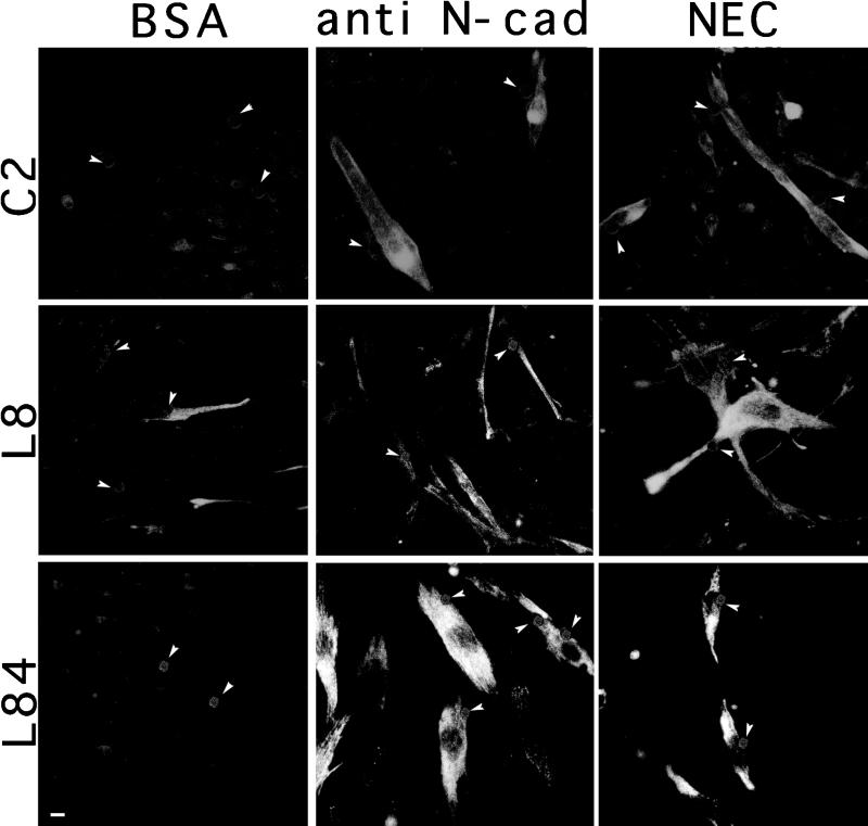
Figure 3.
Expression of skeletal myosin in myocytes treated with beads conjugated to NEC, anti-N-cadherin BE antibodies (anti N-cad) or BSA. C2, L8, and L84 myoblasts were treated with different beads for 48 h, permeabilized, fixed, and immunostained with anti-skeletal myosin antibodies. C2 cells were maintained in growth medium, whereas for L8 and L84 cells growth medium was replaced with differentiation medium simultaneously with the addition of beads. The number of cells per field was approximately equal. Notice the increase in myosin expression in the cultures after treatment with the cadherin-reactive beads. The position of individual beads was detected by phase-contrast microscopy, and their location is indicated by arrowheads. Bar, 10 μm.

