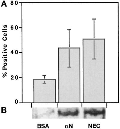
Figure 5.
Effect of cadherin-reactive beads on the expression of skeletal α-actinin in C2 cells. C2 myoblasts were plated and cultured overnight on gelatin-coated coverslips (A) or tissue culture dishes (B) at 50% confluence. Beads were added for 48 h, and then the samples were either fixed and immunostained or subjected to protein extraction and SDS-PAGE immunoblot analysis with antibodies against skeletal α-actinin. (A) Calculation of the percentage of α-actinin–positive, bead-bound cells. Each column represents the mean ± SD of 4 independent experiments; 200 cells were counted in each case. (B) Representative immunoblot analysis of the total cell extracts prepared from C2 cells treated as in A. αN, anti-N-cadherin antibodies.

