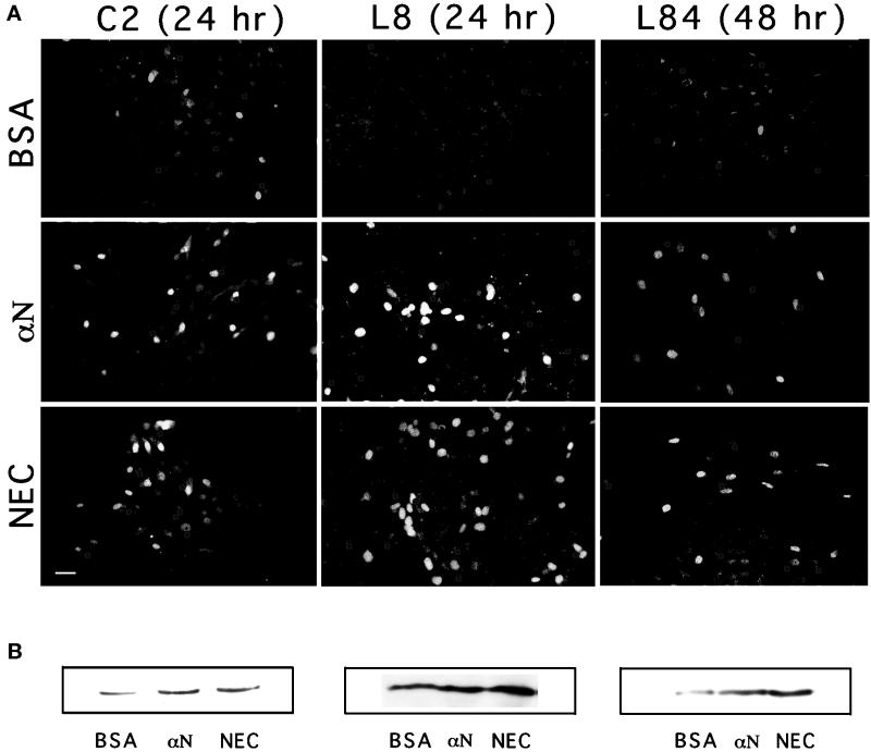
Figure 7.
Effects of N-cadherin stimulation on myogenin expression in cultured myoblasts. C2, L8, or L84 myoblasts were seeded and incubated overnight on gelatin-coated coverslips, treated with different beads, as indicated, permeabilized, fixed, and immunostained with anti-myogenin antibodies (A). The number of cells, as determined by DAPI staining, was approximately equal in all fields, and the percentage of bead-associated cells was ∼25%. Notice the overall increase in the number of myogenin-positive nuclei in cells after treatment with the cadherin-reactive beads. Bar, 20 μm. (B) Immunoblot analysis of total cell extracts prepared from the three myogenic lines, treated as in A. αN, anti-N-cadherin antibodies.

