Figure 1.
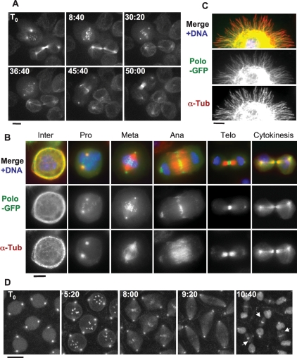
Polo localizes to MTs in a cell cycle-dependent manner. In addition to centrosomes, kinetochores, and the midbody, Polo-GFP localizes to MTs in cytokinesis and interphase. (A) Polo-GFP dynamics in D-Mel cells by time lapse (time is shown in minutes:seconds). Bar, 5 μm. See Supplemental Movie 1 and the text for details. (B) Polo-GFP colocalizes with MTs during interphase and cytokinesis. Cells were fixed with formaldehyde. Polo-GFP appears in green, α-Tubulin is stained in red, and DNA is DAPI-stained in blue. Bar, 5 μm. (C) Polo-GFP colocalizes with MTs in a cell spreading on a concanavalin-A-coated surface and fixed with methanol/formaldehyde (only a portion of the cell is shown for clarity). Stainings are anti-GFP (green), α-Tubulin (red), and DNA (blue). Bar, 5 μm. (D) GFP-Polo dynamics in a transgenic syncytial embryo (cycle 13) by time lapse (time is shown in minutes:seconds). Note that GFP-Polo localizes to the MTs of the central spindle during karyokinesis (arrows). Bar, 10 μm.

