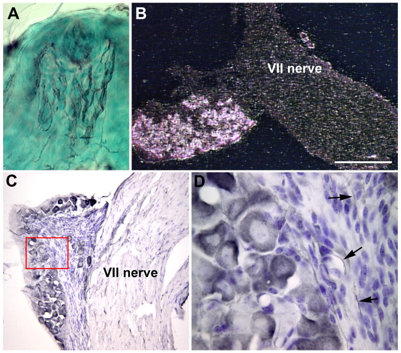
Figure 1.
Gustatory neurons express the p75 receptor. A fungiform papilla (A) labeled with an antibody to p75 demonstrates labeling of nerve fibers in the papilla core as well as in the taste bud (black arrows). A darkfield image of the geniculate ganglion following p75 in situ hybridization (B) illustrates that most neurons in the geniculate ganglion (between arrows) express p75 mRNA. A geniculate ganglion labeled with an antibody to p75 (C) demonstrates that all the ganglion neurons contain this protein although some are more intensely labeled than others. The area outlined by the red box in C is shown at higher magnification (D). p75 labeling can be seen in the cytoplasm of all neurons, but not in the satellite cells. Some p75-labeled fibers can also be identified in the nerve bundles (arrows). Scale bar in B = 250 µm.

