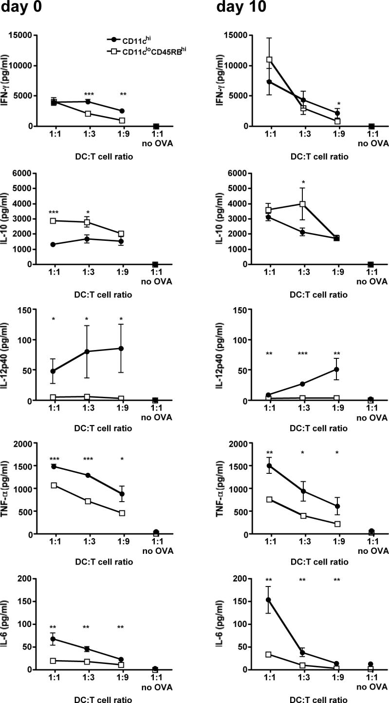
Figure 4. Conventional DCs induce a stronger inflammatory cytokine environment.

Mice were injected i.p. with 106 infected erythrocytes. At days 0 (naive mice) and 10 post-injection, DC subsets were sorted by FACS. DC subsets were then co-cultured at various ratios with naive CD4 T cells isolated from DO11.10 mice, with or without OVA peptide. After 80 hours of co-culture, the culture media was assayed for cytokine content using the BD Cytometric Bead Array. Error bars represent standard deviation within co-cultures of cells sorted from 3 individual mice (*, P < 0.05; **, P<0.01; ***, P < 0.001 when comparing CD11chi co-cultures to CD11cloCD45RBhi co-cultures by Student's t-test).
