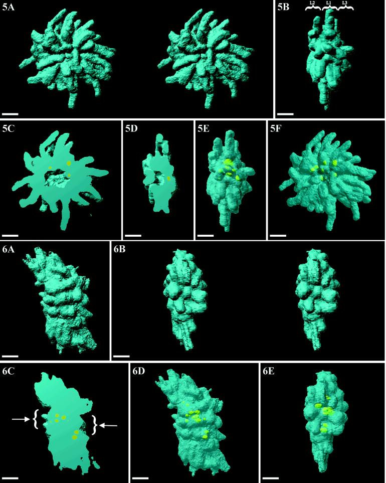
Figure 5.
Volumic visualization of an entire prometaphase cell. The volumic arrangement of the chromosomes (blue) as three layers is revealed in a front-view stereo-pair (A) and a side-on view (B) (L1, L2, and L3). By cutting away half of the volume, the radial disposition of the chromosomes and the central cavity are revealed (C and D). Localization of UBF (yellow) within the NORs appears when DNA is rendered partly transparent: (E) front view; (F) side-on view. Bars, 2 μm.

