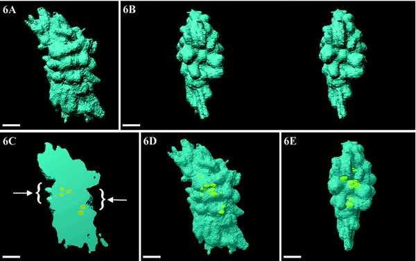
Figure 6.

Volumic visualization of an entire metaphase cell. (A) Chromosomes (blue) are tightly packed and less visible. (B) Stereo-pair of a polar view showing the presence of a depression, facing the pole, which is limited by chromosomes protruding toward the pole. (C) By cutting half of the chromosomes, the two depressions (arrows) and the internal position of NORs are revealed. (D and E) By rendering DNA transparent, the NORs appear clearly as doublets that are aligned in one central plane (E). Bars, 2 μm.
