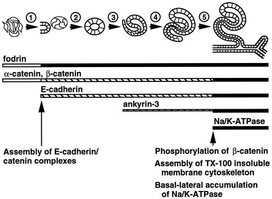
Figure 5.
Model of membrane cytoskeleton assembly and basal-lateral accumulation of Na/K-ATPase in the renal epithelium in situ. The schematic time line at the top depicts different stages of early renal epithelial differentiation (nonpolarized mesenchymal cells, cellular aggregates, renal vesicles, comma-shaped bodies, S-shaped bodies, and early tubules, from left to right). The numbers above the arrows identify different events during renal epithelial differentiation: (1) induction and compaction of mesenchymal cells; (2) initial polarization and lumen formation; (3) elongation of renal vesicles and morphological transformation into comma-shaped bodies; (4) elongation and morphological transformation of comma-shaped bodies into S-shaped bodies; (5) fusion of S-shaped bodies with the ureteric system to form early tubules. Horizontal bars below the schematic time line represent the times at which various proteins are expressed; thicker lines correspond to relatively higher levels of protein expression. Open bars represent diffuse cytosolic expression, striped bars represent apical-lateral membrane expression, and filled bars represent uniform lateral membrane expression. Arrows and text at the bottom indicate times at which important events are thought to occur during development of E-cadherin–mediated cell–cell adhesion and assembly of the membrane cytoskeleton.

