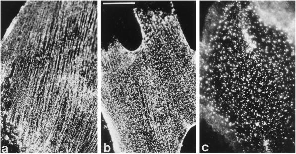
Figure 4.
Cell surface distribution of NG2. Immunofluorescence staining with rabbit antibody against NG2 was used to compare the distribution of wild-type NG2 and NG2 variants on the surface of B28 cells. Cells were allowed to grow overnight on tissue culture dishes coated with PLL to achieve maximal spreading. Immunostaining was carried out at room temperature with living cells. (a) Wild-type NG2 (B28NG2.6). (b) Truncated NG2 (B28NG2/t3.6). (c) Chimeric NG2 (B28NG2/CNTN. M). Bar in b, 10 μm.

