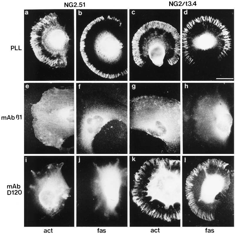
Figure 8.
Distribution of the actin-binding protein fascin during spreading of U251 transfectants. U251NG2.51 (a, b, e, f, i, and j) and U251NG2/t3.4 (c, d, g, h, k, and l) cells were allowed to spread for 40 min on PLL–, mAb β1–, and mAb D120–coated surfaces. Cells were then fixed with methanol and stained with mAbs against either β-actin (act; a, e, i, c, g, and k) or fascin (fas; b, f, j, d, h, and l). On PLL, both cell types contain radial actin spikes decorated with fascin (a–d), whereas on mAb β1, these radial spikes are absent (e–h; note some evidence of stress fiber formation in panel e, although the β-actin antibody is substantially inferior to phalloidin in labeling these structures). On mAb D120 (i–l), radial actin spikes decorated with fascin are seen only in the case of the NG2/t3.4 transfectants. Bar in d, 10 μm.

