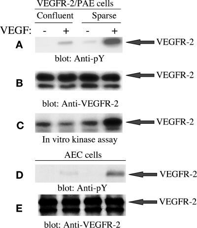
Figure 1.
Effect of endothelial cell density on activation of VEGFR-2. An equal number of PAE cells overexpressing VEGFR-2 or AEC cells endogenously expressing VEGFR-2 were cultured in 10-cm (dense condition) or 15-cm (sparse condition) tissue culture plates, serum starved overnight, and stimulated with VEGF (100 ng/ml) for 5 min. Cells were lysed and immunoprecipitated with an anti-VEGFR-2 antibody and immunoblotted with an anti-phosphotyrosine (pY) antibody (A and D) or subjected to an in vitro kinase assay (C). To determine the protein levels in each lane, the same membranes were reprobed with an anti-VEGFR-2 antibody (B and E).

