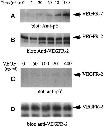
Figure 2.
Kinetics of tyrosine phosphorylation of VEGFR-2 in confluent PAE cells. Serum-starved confluent PAE cells overexpressing VEGFR-2 were stimulated with VEGF (100 ng/ml) for the indicated times (A and B) or with the increasing concentrations of VEGF (C and D). Cells were lysed and immunoprecipitated with an anti-VEGFR-2 antibody. The immunoprecipitated proteins were collected, resolved on SDS-PAGE, transferred to an Immobilon membrane, and immunoblotted with an anti-phosphotyrosine (pY) antibody (A and C). To determine the protein levels in each lane, the same membrane was reprobed with an anti-VEGFR-2 antibody (B and D).

