Figure 2.
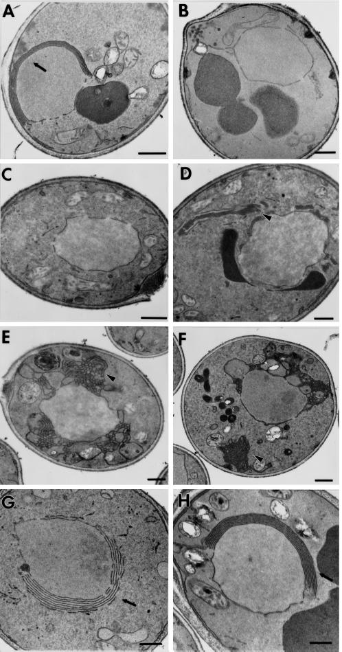
The ultrastructure of the nuclear envelope in a wild-type strain and in strains expressing the Hmg1 fusion proteins was compared. (A) A wild-type strain expressing intact Hmg1p (RWY 410) contains karmellae membranes in an ordered array on the nucleus. (B) Cells expressing the truncated Hmg1:HA protein (RWY 614) lack any ER membrane proliferations. (C and D) Cells expressing Hmg1Δ29p (RWY 406) usually lack karmellae and sometimes produce extended ER networks. (E and F) Cells expressing the Hmg1mem:GFP (RWY 626) do not generate karmellae but have altered ER arrays. (G) Cells expressing the Hmg1mem:Hmg1525–987:GFP (RWY 621) generate karmellae. (H) Cells expressing the Hmg1mem:Hmg2 cat (RWY 572) generate karmellae predominantly. Occasionally, Hmg2-type membranes are found in the population of cells expressing the Hmg1mem:Hmg2cat. Arrows point to karmellae; arrowheads point to proliferated ER. Bars, 0.5 μm.

