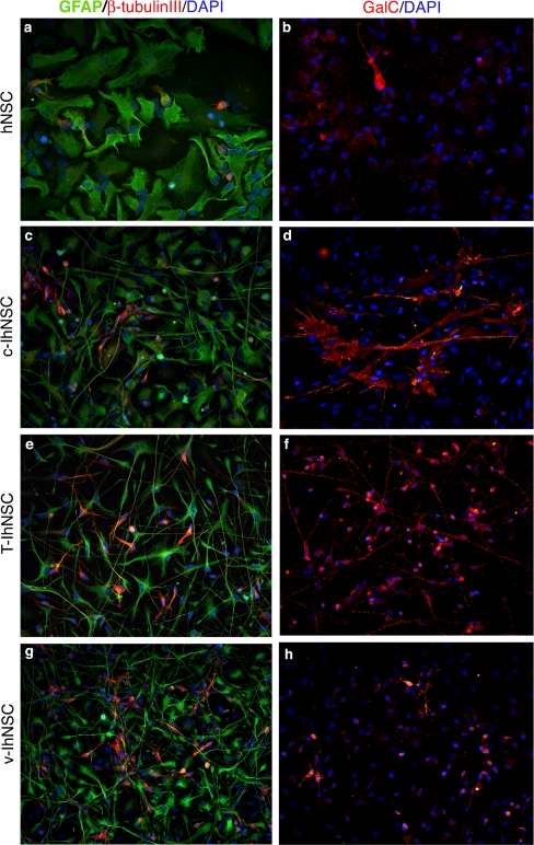
Figure 2. Multipotency.
Cells from hNSC, c-IhNSC, T-IhNSC and v-IhNSC cells line were differentiated on laminin-coated glass coverslips in the absence of growth factors (see materials and methods) for 10 (a,c,e,g) and 17 days (b,d,f,h). Immunofluorescence staining showing the presence of astrocytes (GFAP+, green, a,c,e,g), neurons (β-tubulin+, red, a,c,e,g) and oligodendrocytes (GalC+, red, b,d,f,h). The three major neural lineage markers were detected in distinct cells of the same cell population of origin, thus demonstrating the multipotency of IhNSCs. DAPI nuclear staining (blue) is also shown to detect total cells. Magnification 20×.

