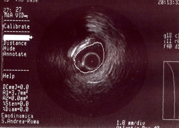
Figure 11.

A-dimensional display mode of IVUS of the same segment as in figure 7. The cross-sectional image shows severe stenosis in the middle segment of the obtuse marginal branch (the residual lumen appears as an anechoic area). Note the calcified tissue and an ulcerative lesion.
