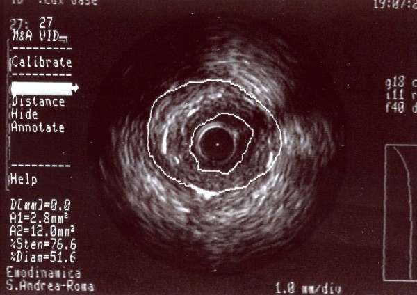
Figure 8.

A-dimensional display mode of IVUS of the same segment as in figure 6. The cross-sectional intrastent image shows severe stenosis in the second segment of the right coronary artery (the residual lumen appears as an anechoic area). Note a thick fibrous cap consisting of fibrous tissue.
