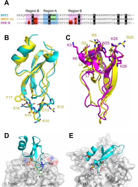
Figure 6. Structural comparison between HWTX-XI with BPTI and DTX-K and docking superimposition of HWTX-XI with Trypsin and pore region of KV1.1.
(A). Sequence alignment of PBTI, DTXK and HWTX-XI. (B). Structural superimposition of HWTX-XI in yellow with PBTI in cyan. (C) Structural superimposition of HWTX-XI in yellow with DTX-K in magentas. (D) Docking superimposition of HWTX-XI with trypsin. (E) Docking superimposition of HWTX-XI with pore region of KV1.1. All key residues of HWTX-XI are represented as stick and coloured in rainbow. Some residues of trypsin and Kv1.1 are presented as sticks and the parts of the surface rounding the pore of the channel are colored red.

