
Fig 3.
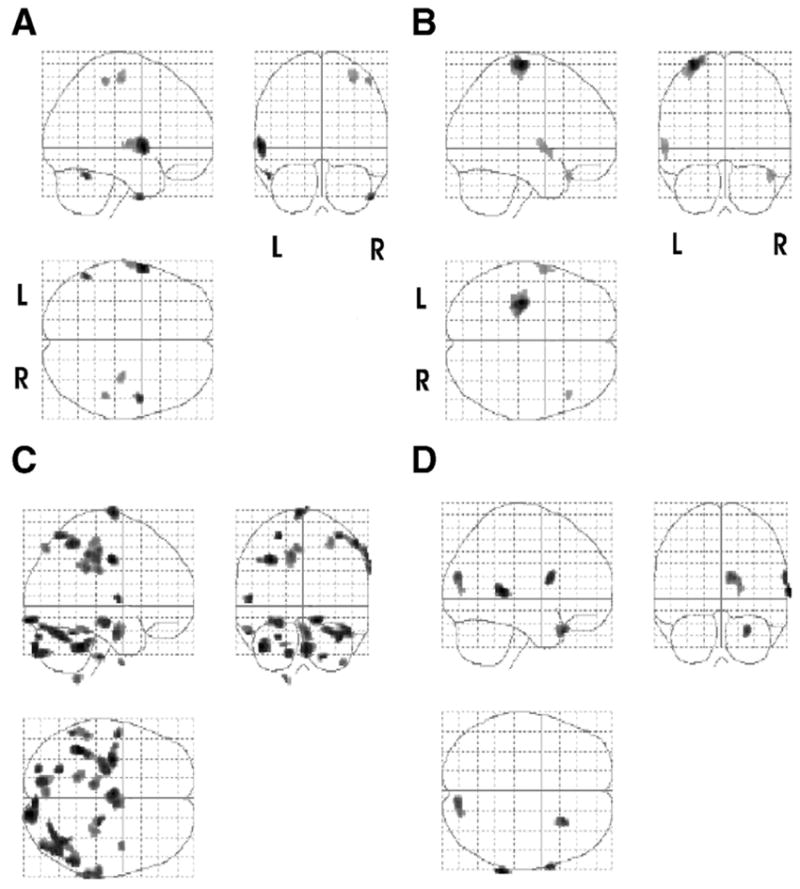
Cortical changes associated CIMT plus mental practice. Images reflecting the activations in 4 subtractions in patient 2. The top row of images depicts the sites of activation by subtracting the rest condition from the actual movement of the affected (right) hand condition (A) pretreatment (move affected > rest) and (B) posttreatment (move affected > rest). The second row depicts the sites from the subtraction of the rest from imagine moving the right hand condition both (C) pretreatment (imagine move affected > rest) and (D) posttreatment (imagine move affected > rest). Note (D) increased ipsilateral cortical activation. Shown are all activations that passed a criterion of P <.05 corrected for multiple comparisons with an extent threshold of 0.