
Fig. 2.
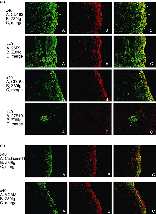
(a) Double-immunofluorescence staining for the Z39Ig antigen and the CD163, 25F9 or CD16 in RA synovium, and for the Z39Ig antigen and 27E10 antigen in the synovial sublining layer of RA synovium. Frozen sections of RA synovium were double-stained with anti-Z39Ig mAb and indicated mAbs, and analysed by the fluorescent microscopy as described in Materials and Methods. The data are representative of independent experiments from four different RA synovium. (b) Double-immunofluorescence staining for the Z39Ig antigen and the cadherin-11 or VCAM-1 antigen in RA synovium. Frozen sections of RA synovium were stained with anti-Z39Ig mAb and anti-cadherin-11 or anti-VCAM-1 mAb, and analysed by fluorescent microscopy as described in Materials and Methods. The data are representative of independent experiments from four different RA synovium.