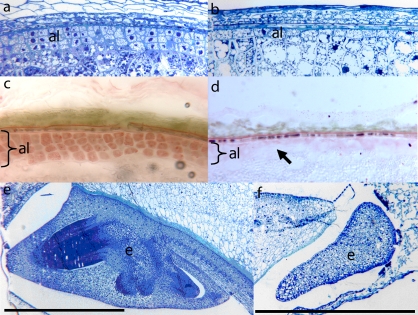
Fig. 1.
Microsections showing the peripheral cell layers of wt (a) and des5 kernels (b). (c, d) Sudan Red stained cryosections of wt (c) and des5 kernels (d). Sudan red is a specific stain for lipid accumulation, and often used as a marker for the lipid-rich aleurone cells. The arrow indicates missing aleurone cell layers in the mutant. (e, f) Microsections showing the embryo of wt (e) and des5 kernels (f). The des5 embryo arrests at an early stage. Al, aleurone; e, embryo. Black scale bars are 1 mm long.

