
Figure 9.
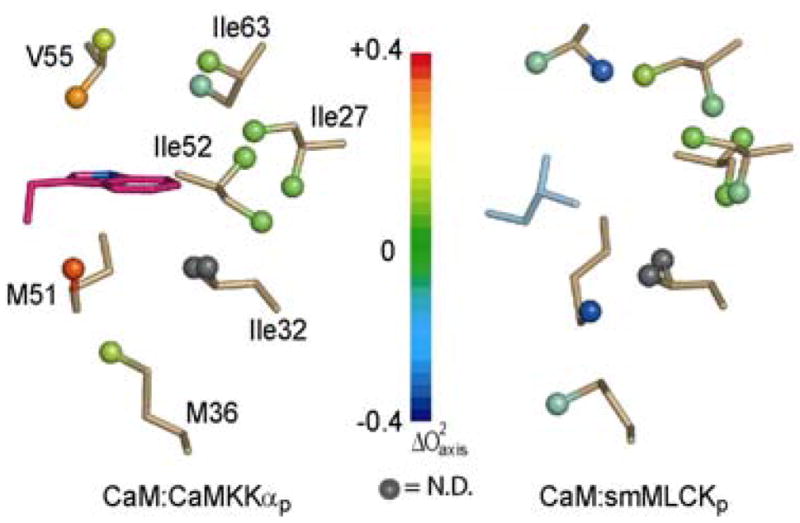
The dynamic effect of different hydrophobic anchors in the amino-terminal EF-hand domain. The two structures were globally superimposed using CaM backbone atoms (2.13Å rmsd). Methyl bearing residues in close contact with the anchor residues are shown in gold sticks with color coded balls for methyl groups. The spatially equivalent residues from each peptide are shown in magenta for W7(CaMKKαp) and cyan for L813(smMLCKp). Color coding reflects changes in values.