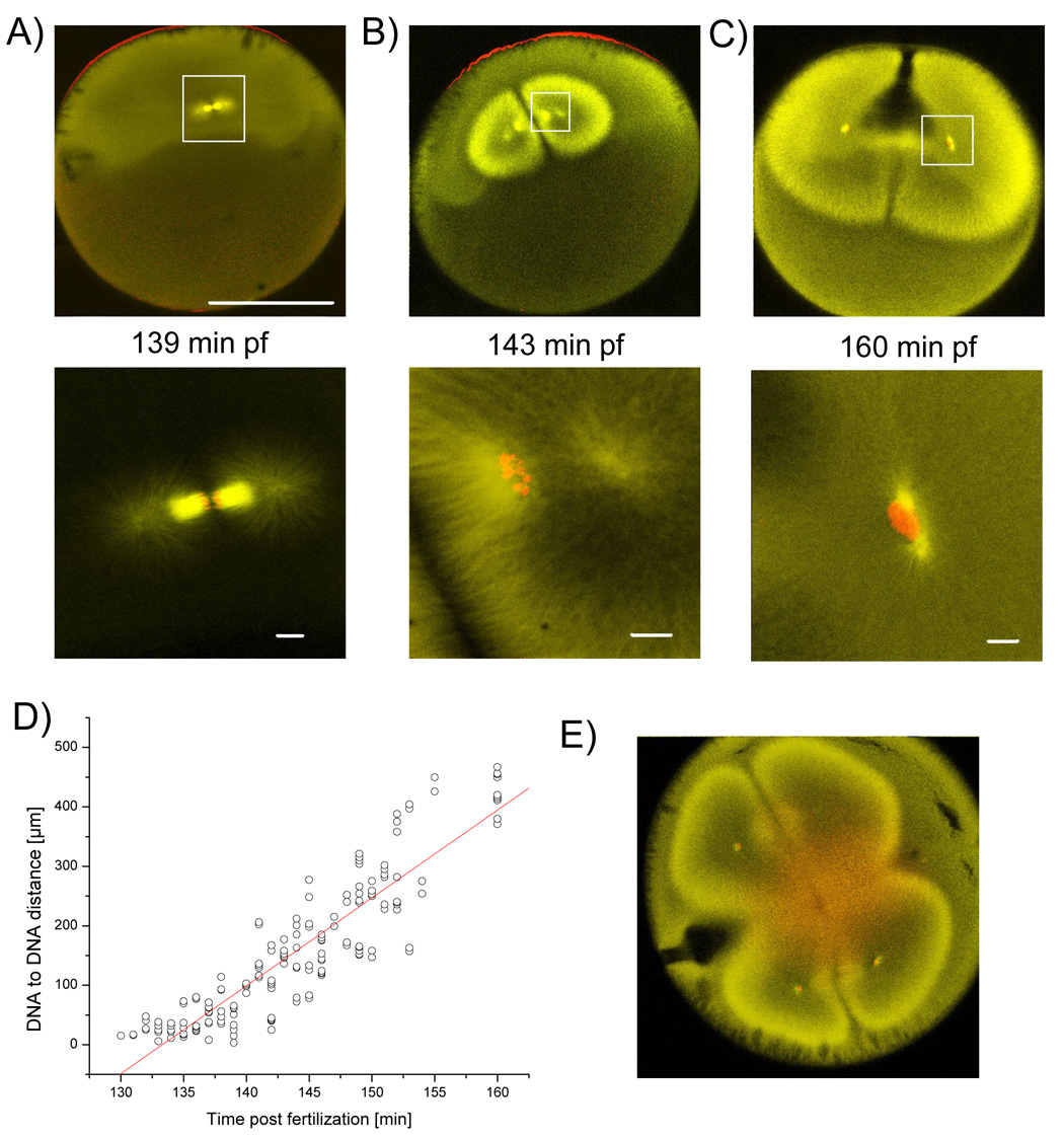
Figure 4. Relatively small spindle is compensated by enormous anaphase B like movement.
Embryos of a synchronously fertilized population were fixed between first and second cytokineses and stained for tubulin (yellow) and DNA (red). A) At anaphase the astral microtubules start to elongate. B) Up to a DNA to DNA distance of ~180 µm, DNA is still condensed and surrounded by high staining of microtubules. Astral microtubules form a hollow structure. C) Nuclear envelope has reformed and finally the nuclei have been separated by ~400 µm, astral microtubules reach the cell cortex and cytokinesis starts. A–C) Bar for upper row = 500 µm, Bars in lower row = 20 µm. D) Plot of DNA to DNA distance versus time. Linear fit estimates speed of DNA separation at ~15 µm/min. E) Cytokinesis, but not separation of DNA, is inhibited by addition of 33 µg/ml of actin depolymerazing Cytochalasin B.

