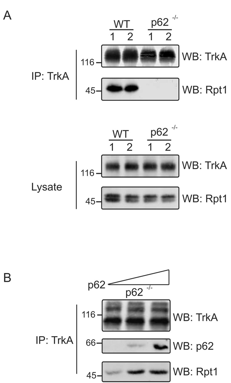
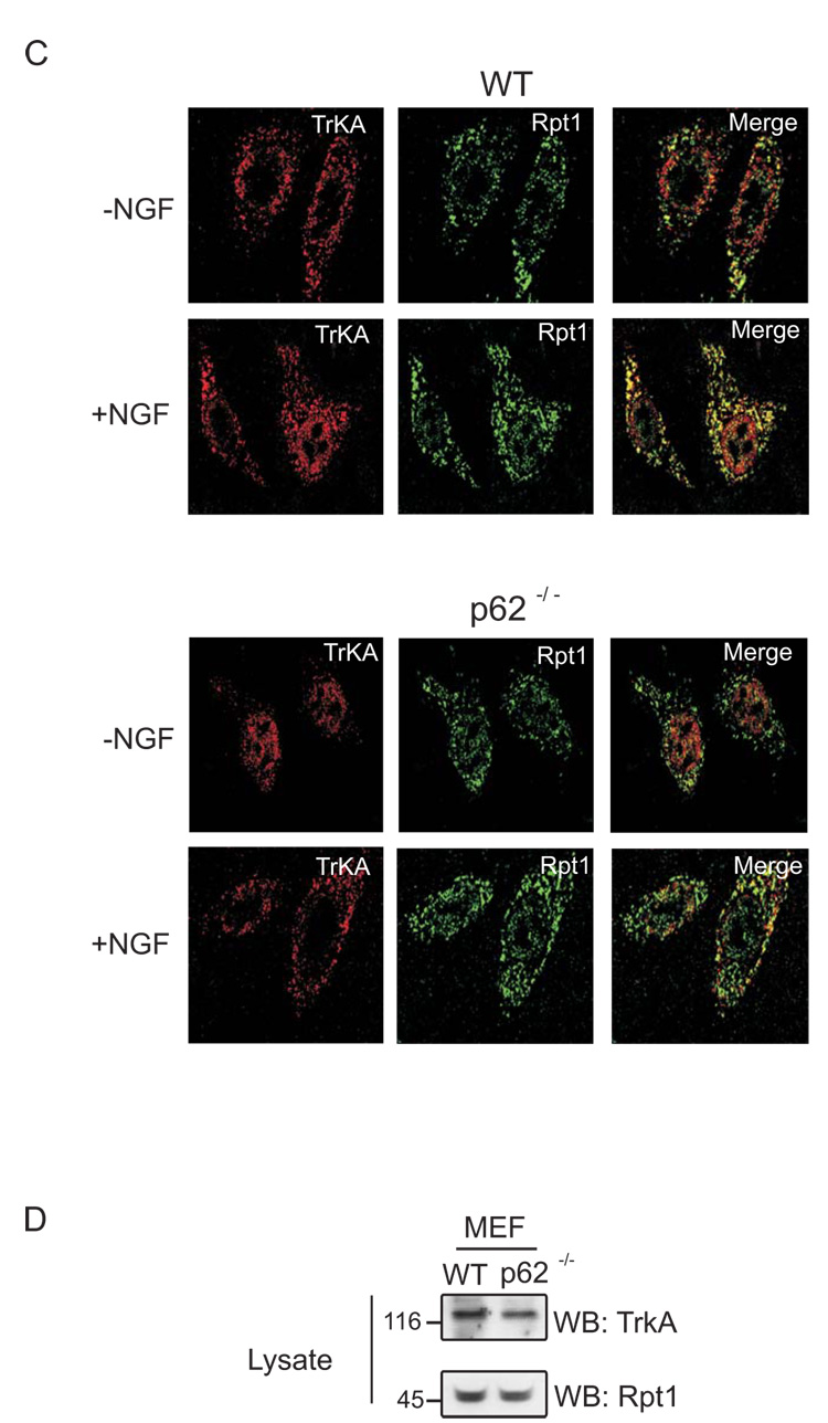
Fig. 1. Interaction of TrkA with Rpt1 is p62 dependent.
(A) Brain of two mice each, WT and p62 −/− were homogenized in Triton lysis buffer. The homogenate was immunoprecipitated (IP) with TrkA (C-14) antibody and Western blotted (WB) with TrkA (B-3) and Rpt1 antibody. (B) Purified p62 (0, 50, 250 ng) was added to isolated proteasomes from p62 −/− mouse brain and rotated for 1 h at room temperature. TrkA was immunoprecipitated (IP) and Western blotted (WB) with TrkA (B-3), p62 and Rpt1 antibody. (C) WT and p62 −/− MEF cells were stimulated with or without NGF (50 ng/ml) for 1 h, followed by fixation and staining with both anti-TrkA (B-3) and anti-Rpt1. The extent of colocalization (Merge, yellow) was assessed by superimposing red (TrkA, Texas Red) and green (Rpt1, Oregon Green) signals. (D) 50 µg of WT and p62 −/− MEF cells were Western blotted (WB) with TrkA (B-3) and Rpt1 to check their expression.


