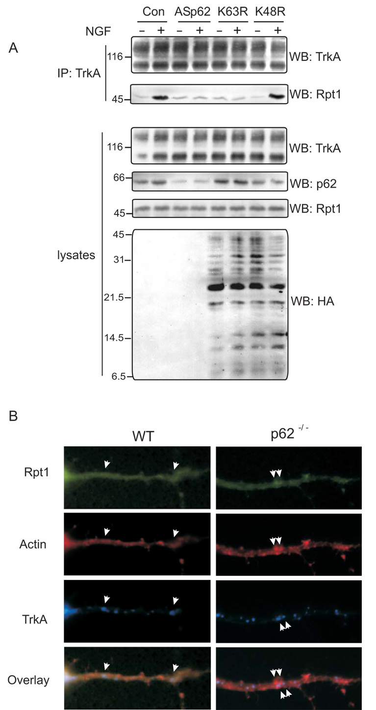
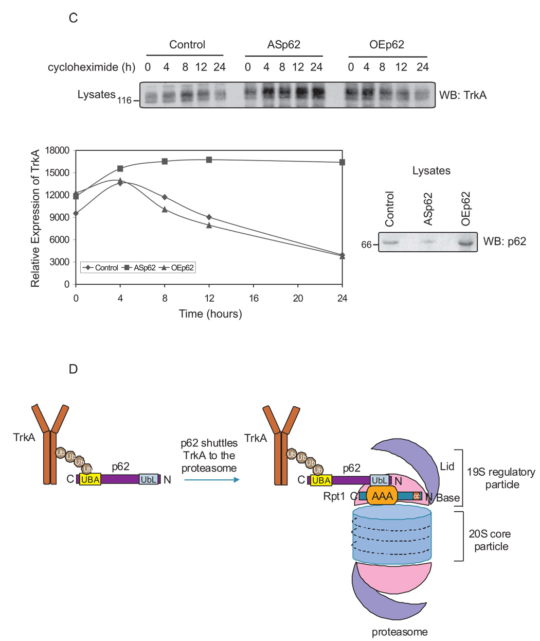
Fig. 4. TrkA turnover is p62 dependent.
(A) PC12 cells were transfected with antisense (AS) p62, HA-tag K63R or K48R mutant ubiquitin constructs. The cells were treated with NGF (50 ng/ml) for 60 min or not before lysing the cells in Triton lysis buffer. Proteins (750 µg) were immunoprecipitated (IP) with anti-TrkA (C-14) and analysed by Western blotting (WB) with TrkA (B-3) and Rpt1 antibody. The expression of TrkA, p62 and ubiquitin constructs in the lysates (50 µg) was determined by Western blotting. (B) Cortico-hippocampal neurons transfected with GFP-Rpt1, HA-TrkA were stimulated with 60 mM K+ for 15 min followed by fixation and staining for actin with phallodin (red), or HA for TrkA. Secondary antibody tagged with CY5 (Molecular Probes, Eugene, OR) was employed to detect HA-TrkA. The merged image is shown. When all three proteins colocalize (red-green-blue) a lavender image appears, whereas miscolocalization of GFP-Rpt1 results in red-blue that produces a violet image. (C) PC12 cells were transfected either with ASp62 or p62 (OEp62). Thirty-six hours post-transfection, the cells were treated with 20 µg of cycloheximide at 37°C for the indicated time periods and lysed with Triton lysis buffer. Equal amounts of cell lysate (50 µg) were Western blotted (WB) with anti-TrkA (B-3) to assess the turnover of endogenous TrkA. The TrkA blot was scanned and the relative expression of TrkA is represented in the graphical plot. The lysates were analyzed for protein expression of p62. (D) Schematic representation of TrkA/p62 proteasomal shuttling. Ubiquitinated TrkA [2] interacts with p62's UBA domain [10] and is targeted by the UbL domain of p62 to the proteasome by interacting with Rpt1. These findings (A–C) are representative of three other independent experiments.


