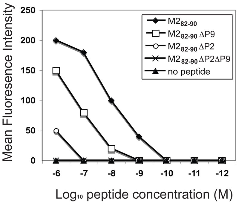
Figure 1.
Binding of wild-type peptides or peptides mutated at H2-Kd anchor residues. RMA-S-Kd cells were incubated at 26 °C, and mixed with peptides at various concentrations. Binding of M282–90 (SYIGSINNI) or mutated peptides, M282–90ΔP2 (SRIGSINNI), M282–90ΔP9 (SYIGSINNT), and M282–90ΔP2ΔP9 (SRIGSINNT) to H2-Kd molecules was compared with that of the no-peptide control. The mean fluorescence intensity determined by FACS analysis is plotted against peptide concentration. The results shown represent the data from a single representative experiment, which was one of three with similar results.

