
Figure 4. Cytoskeletal reorganization and formation of actin stress fibers during the switch from dormancy to metastatic growth.
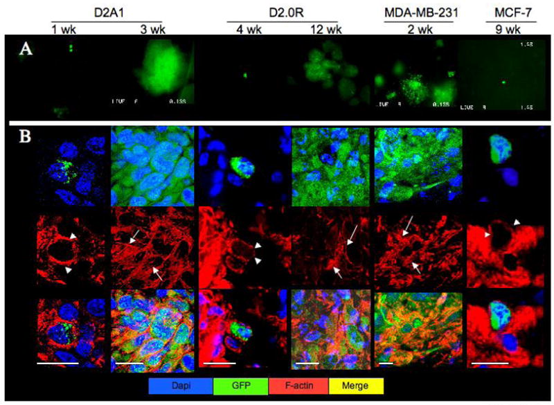
A) SCOM images (magnification x100) of lungs from mice injected with either D2A1-GFP (removed 1 or 3 weeks post-injection), D2.0R GFP (removed 4 or 12 weeks post-injection) MDA-MB-231-GFP (removed 2 weeks post-injection) or MCF-7-GFP cells (removed 9 weeks post-injection). Time points varied according to the growth properties of the cells. B) Frozen sections of lungs from mice injected with the above cells at the indicated time points stained for f-actin (red); confocal microscopy, white bar equals 20 microns. White arrowheads depict cortical actin staining; white arrows indicate f-actin stress fibers.